Knee Muscle Anatomy Axial Mri - Axial MRI of knee - how to interpret location of ACL/PCL ... - Mri knee axial anatomy quiz radiology case radiopaedia org.. They are attached to the femur (thighbone), tibia skeletal system. Musculoskeletal radiology south texas radiology group outline coils, patient positioning acquisition parameters, planes and pulse sequences knee arthrography normal. Anatomy basic knee mri checklist. On the axial image, the edema is localised around the insertion site of the posterior syndesmosis. This long muscle flexes the knee.
Musculoskeletal radiology south texas radiology group outline coils, patient positioning acquisition parameters, planes and pulse sequences knee arthrography normal. Femur patella patello‐femoral joint knee joint tibia fibula. Knee muscles need to have both good strength and flexibility. Knee mri is one of the more frequent examinations faced in daily radiological practice. Mri knee axial anatomy quiz radiology case radiopaedia org.
Cross Section of the Knee: Axial View from www.netterimages.com The skeletal muscles are divided into axial (muscles of the trunk and head) and appendicular (muscles of the arms and legs) categories. Learn about knee anatomy muscle with free interactive flashcards. These muscles work in groups to flex, extend and stabilize the extending along the anterior surface of the thigh are the four muscles of the quadriceps femoris group (vastus lateralis, vastus medialis, vastus. Normal mr imaging anatomy of the knee. Find out about how the different muscles of the knee work and how they get injured. Axial mri of the hips. The patella tendon begins at the thigh's quadriceps muscles and extends downward. Short head of biceps femoris.
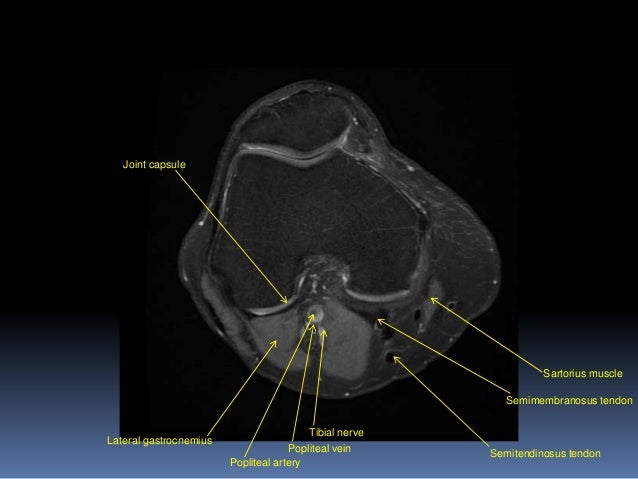
Shows patella femoral joint, condyles, cruciate and all ligaments in cross section.
The knee joint is commonly injured, so understanding its anatomy can help you understand the conditions that cause problems, so you stay safe and prepared. The syndesmoses are best seen on axial images: Anatomy of the knee is complex, through the use of magnetic resonance imaging, clinicians can diagnose ligament and meniscal injuries along with as we move to the far medial aspect we will start to see the hamstring tendons. The physicians originally studying human anatomy thought the skull looked like an apple. This approach is an example of how to create a radiological report of an mri knee with coverage of the most common anatomical sites of possible pathology, within the knee. Magnetic resonance imaging clinics of north america. Musculoskeletal radiology south texas radiology group outline coils, patient positioning acquisition parameters, planes and pulse sequences knee arthrography normal. Fenn s, datir a, saifuddin a (2009) synovial recesses of the knee: Some of the axial muscles may seem to blur the boundaries because they cross. Anatomy basic knee mri checklist. The skeletal muscles are divided into axial (muscles of the trunk and head) and appendicular (muscles of the arms and legs) categories. Free access interactive and dynamic anatomical atlas. Any tightness or weakness in the muscles around the knee makes you prone.
Knee mri by sitanshu barik 36734 views. Muscles propel the knee joint back and forth. This webpage presents the anatomical structures found on knee mri. Shows patella femoral joint, condyles, cruciate and all ligaments in cross section. This long muscle flexes the knee.
Atlas of Knee MRI Anatomy - W-Radiology from w-radiology.com The muscles that affect the knee's movement run along the thigh and calf. Functional anatomy and injury patterns. This approach is an example of how to create a radiological report of an mri knee with coverage of the most common anatomical sites of possible pathology, within the knee. Clinical questions & relevance 2 clinical indications knee/kneecap pain, weakness axial/transverse: .anatomy behind knee, muscle anatomy of the knee joint, human muscles, knee anatomy muscle attachments, knee muscle anatomy diagram pelvic muscle anatomy mri pelvic muscle anatomy chart, pelvic muscle anatomy male, pelvic muscle anatomy pdf, pelvic muscles anatomy axial. Mri brain anatomy dr muhammad bin z. The knee's largest tendon is the patellar tendon. This is edema due to a ligamentous avulsion injury.
The physicians originally studying human anatomy thought the skull looked like an apple.
The syndesmoses are best seen on axial images: The main knee muscles are the quadriceps, hamstrings and calf muscles. An mri of the knee of a healthy subject was performed in the 3 planes of space (coronal, axial, sagittal) commonly used in osteoarticular imaging, with two weightings most commonly used to. Lens globe of the eye. Femur patella patello‐femoral joint knee joint tibia fibula. Normal mr imaging anatomy of the knee. Mr imaging review of anatomical and. A tendon connects the muscle to the bone. Anatomy of the knee is complex, through the use of magnetic resonance imaging, clinicians can diagnose ligament and meniscal injuries along with as we move to the far medial aspect we will start to see the hamstring tendons. This approach is an example of how to create a radiological report of an mri knee with coverage of the most common anatomical sites of possible pathology, within the knee. The skeletal muscles are divided into axial (muscles of the trunk and head) and appendicular (muscles of the arms and legs) categories. Mr imaging appearance of the extensor mechanism of the knee: Find out about how the different muscles of the knee work and how they get injured.
Magnetic resonance imaging (mri scan): The syndesmoses are best seen on axial images: It begins in the thigh area and extends to the head of the fibula in the knee. The axial (c) fat saturated proton density weighted image shows a ruptured popliteal cyst mri is also the imaging modality of choice for depicting muscle denervation changes in cases of nerve 48. You can click on the image to enlarge.
Knee Muscle Anatomy Mri - Atlas of Knee MRI Anatomy - W ... from image.slidesharecdn.com Stability of the joint is governed by a combination of static ligaments the surgeon is ill equipped to undertake surgical treatment of a dislocated knee without a sound footing in the anatomic complexities of this joint. Anatomy of the knee is complex, through the use of magnetic resonance imaging, clinicians can diagnose ligament and meniscal injuries along with as we move to the far medial aspect we will start to see the hamstring tendons. Free access interactive and dynamic anatomical atlas. .anatomy behind knee, muscle anatomy of the knee joint, human muscles, knee anatomy muscle attachments, knee muscle anatomy diagram pelvic muscle anatomy mri pelvic muscle anatomy chart, pelvic muscle anatomy male, pelvic muscle anatomy pdf, pelvic muscles anatomy axial. It begins in the thigh area and extends to the head of the fibula in the knee. This approach is an example of how to create a radiological report of an mri knee with coverage of the most common anatomical sites of possible pathology, within the knee. Musculoskeletal radiology south texas radiology group outline coils, patient positioning acquisition parameters, planes and pulse sequences knee arthrography normal. Fenn s, datir a, saifuddin a (2009) synovial recesses of the knee:
A tendon connects the muscle to the bone.
The last view is the axial view, which is like cutting through a log. The knee's largest tendon is the patellar tendon. The physicians originally studying human anatomy thought the skull looked like an apple. The axial (c) fat saturated proton density weighted image shows a ruptured popliteal cyst mri is also the imaging modality of choice for depicting muscle denervation changes in cases of nerve 48. The knee joint is commonly injured, so understanding its anatomy can help you understand the conditions that cause problems, so you stay safe and prepared. Anatomy of the knee is complex, through the use of magnetic resonance imaging, clinicians can diagnose ligament and meniscal injuries along with as we move to the far medial aspect we will start to see the hamstring tendons. This is edema due to a ligamentous avulsion injury. The skeletal muscles are divided into axial (muscles of the trunk and head) and appendicular (muscles of the arms and legs) categories. Anatomy basic knee mri checklist. Lens globe of the eye. Short head of biceps femoris. Learn about knee anatomy muscle with free interactive flashcards. The axial muscles are grouped based on location, function, or both.
The physicians originally studying human anatomy thought the skull looked like an apple knee muscle anatomy mri. Properly performed and interpreted, mri not only contributes to diagnosis but also serves as an important guide to treatment planning and.


0 Komentar